A new device employing vortex ultrasound aims to transform pulmonary embolism treatment, promising faster clot dissolution and safer procedures.
Introduction to Vortex Ultrasound Technology
This initiative focuses on accelerating and improving the treatment of pulmonary embolism, a condition where blood clots block vital oxygen exchange in the lungs. Pulmonary embolisms can lead to severe complications or even death if not treated promptly. A collaborative effort led by researchers at the University of Michigan is developing a device that uses vortex ultrasound technology, which could represent a shift in treatment paradigms. This minimally invasive device aims to dissolve lethal blood clots quickly and safely, circumventing existing treatment risks and potentially leading to a more effective emergency response for affected patients.
The Mechanism Behind the Device
The innovative device is equipped with a specialized ultrasound source located at the tip of a catheter. By delivering this technology directly to the clot within the blood vessels, the device unleashes swirling sound waves designed to disrupt the clot’s internal structure without harming the surrounding blood vessel walls. This mechanism is particularly noteworthy; it reportedly generates microbubbles within the clot, which contributes to its rapid fragmentation. This localized approach could lessen the chances of collateral damage to healthy tissues, a common concern in existing treatment models. It's a clever blend of physics and medicine, merging wave dynamics with biological responses to facilitate a more effective clot dissolution.
Significance of Addressing Pulmonary Embolism
Pulmonary embolism ranks as the third-leading cause of cardiovascular-related deaths in the U.S., following heart attacks and strokes. The condition primarily arises when clots dislodge from the legs and travel to the lungs, critically obstructing blood flow and exerting lethal pressure on the heart. Despite advancements in medical technology, current emergency interventions come with significant downsides. Clot-busting drugs can take up to 24 hours to show efficacy and often fail in roughly one-third of cases, posing a risk of serious internal bleeding. Mechanical removal, while sometimes necessary, carries the risk of injuring vulnerable blood vessel walls, and severe clots may even require invasive open-chest surgery. The prevalence of this condition indicates an urgent need for more efficient and safer solutions. Redesigning treatment mechanisms is essential, as the current methods leave much to be desired in terms of speed and safety.
Advantages of Vortex Ultrasound
While traditional external ultrasound methods are generally perceived as safer for vessel integrity, they often struggle with accurate targeting and efficacy, particularly in lung tissues where internal structures complicate delivery. The U-M team’s approach shifts the administration of ultrasound directly into the blood vessels, potentially enabling faster treatment and a reduced risk profile for patients. Early indications suggest that this novel method may clear clots in as little as 30 minutes—a significant improvement over existing strategies. These quicker interventions could not only save lives but also reduce the burden on healthcare systems by lowering hospital stays and recovery times. (And this is the part most people overlook: the economic implications of faster treatments could ripple throughout the healthcare industry.)
Funding and Project Timeline
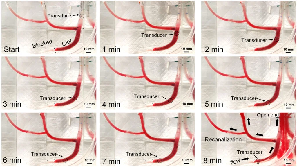
The National Institutes of Health has allocated approximately $3.74 million to support the project over the next five years. This funding is crucial for advancing the research and development of the device. Early tests have shown that the vortex ultrasound can restore blood flow through a simulated three-inch-long clot within just eight minutes, providing promising initial results. These outcomes underline the technology's potential efficacy, but it's worth remembering that rigorous testing lies ahead. The allocated budget will facilitate further refinement of the catheter design, enhancement of the ultrasound wave's shape and frequency, and comprehensive safety evaluations using animal models in preparation for human trials. This structured approach aims to ensure that the technology is not only effective but also safe for clinical application.
Key Contributors
Chengzhi Shi, the principal investigator and associate professor of mechanical engineering at the University of Michigan, underscores the project's ambitious goal: “With this approach, we hope to treat clots in the lungs more quickly and safely while potentially providing an alternative to open-chest surgery for severe pulmonary embolism.” His vision aligns with the pressing need for effective solutions in a high-stakes medical landscape. Collaborating closely with him are several esteemed colleagues, including Daniel Myers and Andrea Obi from the U-M Medical School, along with Zhen Xu from the Department of Biomedical Engineering. Additionally, contributions from Xiaoning Jiang at North Carolina State University highlight a strong interdisciplinary effort. This collaboration enhances the potential for innovative breakthroughs, merging insights from engineering, medicine, and healthcare research.
Implications and Future Outlook
The potential of vortex ultrasound technology could redefine how pulmonary embolism is treated, promising quicker interventions and significant improvements in patient outcomes. Should the subsequent trials prove successful, the implications for emergency medical care could be profound. Healthcare providers will need to reevaluate existing protocols for managing pulmonary embolism, perhaps shifting towards more proactive and technology-driven approaches. The journey from concept to clinical application is well underway, and the results could reshape not only treatment modalities but also patient management strategies in emergency settings. If you're working in this space, you'd be wise to keep a close eye on this development; it could signal the beginning of a new standard in pulmonary care.



Discussion
Sign in to join the discussion.